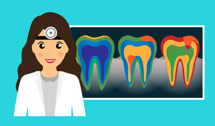
The State of the Art in Dental Image Analysis


Related topics:
Last updated:
Dental image analysis is one of the most challenging and recent areas of medical image analysis. The maxillofacial area is densely populated by complex overlapping structures, often very small. Besides that, teeth significantly vary in shape and position, creating additional obstacles for image segmentation. As a result, dental image analysis requires advanced feature extraction and segmentation techniques, and, in many cases, a 3D reconstruction.
Computer vision applications in dentistry
Medical image analysis mostly deals with diagnostic issues, supporting diagnosticians in their decisions; however, dentistry provides at least one more application for some image analysis techniques, which is computer-aided design and computer-aided manufacturing (CAD/CAM) systems.
Of course, CAD/CAM systems employ only some of image analysis techniques, such as image preprocessing and semi-automated image segmentation, but these systems constitute one of the most important trends in modern dentistry, and this apparently makes them one of the top dental image analysis applications.
Yet, computer-aided diagnostics is the second major use of dental image analysis. Computer vision can be utilized to identify carious lesions and periodontitis signs, as well as maxillary sinusitis, osteoporosis, and other pathologies.
Computer vision is also applied to cephalometric analysis and teeth classification for both theoretical and practical purposes.
CAD/CAM systems
Introduced in the 1980s as an awkward novelty, CAD/CAM systems have since then matured into a cutting-edge orthodontic and dental surgery technique providing a fast and reliable method for dental restoration.
CAD/CAM systems use intraoral cameras or CBCT to reconstruct a 3D picture of the dental arch and provide the information necessary to design and create dental prostheses and orthodontic appliances. The CAM part means that the system creates necessary prostheses on the spot, allowing the manipulation to be performed in a single day.
Global dental CAD/CAM market is consistently growing at an estimated CAGR of over 9% by 2032, and a key trend to foster its growth is the shift toward open architecture software that allows more flexibility.
Diagnostics
As the most common tool based on medical image analysis, computer-aided diagnosis (CAD)[1] systems support radiologists and dentists in their work, often significantly enhancing diagnostic accuracy and speed.
1) Caries
Based on reference matching, CAD systems are already used for caries diagnostics. Some researchers[1] have reported the true-positive result rate to nearly double (from 39% to 69%) in caries detection when supported by a CAD system. Another research[2] has shown less optimistic results due to teeth segmentation problems.
Anyway, carious lesion identification is one of the most common tasks in conservative dentistry, and a partial automation of this process would be of great benefit for physicians, so this problem definitely requires more attention.
2) Endodontic diagnosis
Presence or absence of inflammatory changes in the dental pulp is crucial for the tooth treatment plan. To decide on tooth depulpation, dentists also can ask for CAD system advice.
Several recent research papers propose many-sided approaches to the problem of pulp and root canal segmentation, such as linear prediction model for root canal edge extraction and fuzzy c-means clustering for root canal segmentation in high-resolution micro-CT records.
3) Periodontitis
Periodontitis, an inflammatory disease of soft tissues surrounding the teeth, can be even trickier to detect in plain dental radiographs. In order to succeed, highly sensitive edge detection and segmentation techniques should be applied.
To address the problem of automated periodontitis detection, Chinese researchers suggested a combination of semi-supervised fuzzy clustering algorithms for dental X-ray image segmentation. The proposed system performed much better than conventional segmentation techniques, although the authors admit that there is still room for improvement.
4) Maxillary sinusitis
Dental infections often cause inflammation of maxillary sinuses due to anatomical peculiarities of this area, and it is important that even inexperienced dentists should be able to detect this complication in plain panoramic radiographs. It isn't easy, as dental panoramic images are not intended to diagnose sinus pathology, and overlapping structures often make it difficult to recognize sinusitis.
Research conducted in Japan in 2016 showed that CAD systems make a difference in this case, raising the diagnostic accuracy of neophyte dentists to that of their experienced colleagues. After the initial image filtering and segmentation, the system performs registration of the mirrored sinus regions to detect the difference in the opacity. Although this technique can be applied only to unilateral sinusitis diagnostics, it is still a promising direction.
5) Osteoporosis
While the golden standard for osteoporosis detection is dual energy X-ray absorptiometry (DXA), bone mineral density can also be measured in the area of the mandibular condyle (a part of the lower jaw). Since the dental panoramic radiography is much easier to access than DXA, many doctors see it as a promising method to screen for osteoporosis.
A recent research has shown that using image analysis techniques to determine the ROIs, extract features, and classify the data obtained from panoramic radiographs, it is possible to achieve at least an 88% accuracy rate in osteoporosis detection.
6) Other
Although carotid artery calcification isn't immediately connected with dentistry, it can be spotted in the dental panoramic radiographs. Defining two more ROIs (for both sides) in a panoramic X-ray, an image analysis system can easily detect the calcification with image filtering and grayscale thresholding.[3]
This 'incidental finding' is of vital importance, as it indicates that the patient is at high risk of stroke, so adding such a feature into a CAD system for panoramic X-ray analysis would bring immense benefit.
Cephalometric analysis
Dentists, orthodontist, and oral-maxillofacial surgeons analyze head anatomical landmarks when planning treatment. There is even broader interest in this area, as cephalometry can also be used for various purposes, for example, to plan labor and delivery in obstetrics.
However, manual cephalometric analysis takes significant time, which makes it a good candidate for automation. Medical image analysis systems can drastically increase the speed and efficiency of anatomical landmark detection.
Main challenges
According to a recent survey[2] conducted by an international collaboration, automated detection of carious lesions in bitewing X-ray images is still a big challenge. The key problem is segmentation of dental structures, because the data variation is high, and teeth are sometimes mislabeled as the background.
Another significant challenge outlined in the same paper considers cephalometric analysis. Although the accuracy of automated anatomical landmark detection has increased in the last 7 years, it is still below 90%.
With further development of image processing and machine learning algorithms, the success rate of both caries detection and cephalometric analysis will be increasing.
Trends and growth options
Since CAD/CAM systems are among the key trends in dentistry worldwide, the demand for image preprocessing techniques and segmentation algorithms will grow. And the shift towards open architecture indicates that it will be easier for CAD/CAM manufacturers to order the software tailored for their specific needs.
Speaking about imaging techniques, the palm of victory belongs to cone beam computed tomography, which is another key trend in dentistry. In the nearest future, CBCT empowered with image analysis CAD systems may become the most efficient diagnostic tool in dentistry.
Besides that, the worldwide trend for artificial neural networks, fuzzy logic systems, random forests and other intelligent algorithms that can be used for image clustering and classification creates new opportunities for dental image analysis. Further enhancement of their performance will improve the accuracy of CAD systems and allow automation of the most challenging cases.
1.Tracy KD, Dykstra BA, Gakenheimer DC, Scheetz JP, Lacina S, Scarfe WC, et al. Utility and effectiveness of computer-aided diagnosis of dental caries. General dentistry (2011).
2.C.-W. Wang et al. A benchmark for comparison of dental radiography analysis algorithms. Medical Image Analysis 31 (2016) 63'76.
3.T. Sawagashira, T. Hayashi, T. Hara, A. Katsumata, C. Muramatsu, X. Zhou, et al. An automatic detection method for carotid artery calcifications using top-hat filter on dental panoramic radiographs. IEICE Trans Inform Syst, E96-D (8) (2013), pp. 1878'1881
[1] To avoid ambiguity, we will stress that CAD/CAM stands for computer-aided design and computer-aided manufacturing, while CAD refers to computer-aided detection and diagnosis.
