Automatic brain tumor localization: Expectations and reality


Related topics:
Last updated:
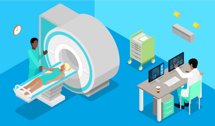
Precise brain tumor localization lies in the correct interpretation of a patient’s brain scans across modalities (MRI, PET, etc.), which allows for a well-timed confirmation of diagnosis. While healthcare is no doubt based on human proficiency and experience, technology appears to be advanced enough to back up health specialists’ decisions with complex mathematical algorithms.
Major advances in creating artificial intelligence solutions with machine learning seem promising to enable automation of medical image analysis, thus reducing time on image interpretation and precisely localizing tumors and their subregions for further treatment prescription and surgery planning.
However, there are many doubts on the reliability of automated localization of brain tumors in the light of multiple challenges on the way. We’ve decided to put together these challenges, define the ways to increase the accuracy of diagnosing tumors in the brain automatically, and also take a look at the state of the art in this field.
The challenges of brain tumor localization
In automated brain tumor analysis, segmentation and registration are the methods that appear most challenging. Some of the following issues remain unsolved, and others can be addressed with a different approach to previous steps (e.g., pre-processing).
Segmentation challenges
Segmentation allows spotting the tumor area, including its sub-compartments and surrounding tissues. Segmenting allegedly tumorous brain images is challenging on several levels:
- If present, high-grade tumors frequently have unclear outlines with fractures. Some tumors can also deform surrounding tissues and come with edema or necrosis, changing the image intensity around the abnormality. This change can hinder accurate localization of the tumor outline, fogging and smudging its borders.
- Single-modality analysis can be insufficient for separating tumor subregions. Therefore, the combination of several modalities might be required to ensure a comprehensive diagnosis. In this case, segmentation will highly depend on the accurate pre-processing and registration stages.
- Full contrast imbibition time and image acquisition time upon contrast injection may differ, which leads to significant changes in tumor appearance. It is still debatable whether there is a need to handle the lesion’s non-imageable component by segmentation algorithms, and if yes, how to achieve that.
Registration challenges
In brain tumor localization, the aim of registration is to enable either simultaneous analysis of different modalities at the diagnostic stage or further monitoring of tumor growth.
A few challenges arise on the way to seamless registration:
- Anisotropic voxel spacing. Images within particular modalities are usually acquired with different anisotropic resolutions, sometimes in different orientations.
- When a patient’s image is registered with a healthy brain atlas, the challenge of missing correspondence between these two arises.
While there is a range of suggested methods to overcome the issues above, most of them require a significant computational time and capacity. This can become one of the major pitfalls on the way to solving segmentation and registration challenges.
Efficient approaches to brain tumor localization
MRI-only diagnostics
To improve tumor localization accuracy in MRI image analysis, a few sequences can be registered. The difference in imaging outcomes of T1 and T2 sequences can ensure precise automatic detection of lesions and their subregions.
For example, a T1-weighted image allows for proper segmentation and, therefore, detects an active tumor and necrosis regions, while the edema region can be segmented based on a registered T2-weighted image. When those two sequences are fused, the image analysis software algorithm is able to form a tumor’s complete overview with all the affected areas.
Cross-modality diagnostics: MRI and PET
Combined with the MRI scan, PET metabolic data (blood flow, oxygen, and glucose metabolism) allows for the creation of a precise picture of how the tumor looks, how it is outlined, and how it affects surrounding tissue, separating the abnormality itself from edema and necrosis.
With high-grade gliomas, for example, the affected area can be misleadingly vast. But when the MRI and PET images are fused, the real division of subregions appears.
State of the art in automated brain tumor localization
Currently, there is a variety of approaches to handle segmentation and registration as primary steps in automating brain cancer diagnostics. But they are rather taken as a set of methods where they can be partially, semi-, or fully automated.
MRI-based medical image analysis for brain tumor studies
In the survey by S. Bauer et al., the authors summarize various sets of approaches to segmentation and registration of brain MRI images. Some of them can be automated, such as:
Segmentation: fuzzy clustering plus knowledge-based techniques, SVM classification, difference image for volumetric tumor assessment, decision forests for tissue-specific segmentation, and more.
Registration: non-rigid registration to capture brain shift, geometric metamorphosis, differential analysis for tumor growth quantification, registration with EM algorithm and diffusion modeling, and more.
But these are only parts that don’t really present an end-to-end system of automatic lesion localization.
Automatic brain tumor segmentation via 3D convolutional neural networks
In 2016, a paper from Stanford University presented a new algorithm for fully automatic brain tumor segmentation based on 3D convolutional neural networks (CNN). The authors (C. Elamri and T. de Planque) claim that this algorithm allows achieving 89% accuracy in the entire tumor segmentation. Researchers also compared their 3D CNN method to the performance of human radiologists (85%), the leading methods of 2013 (75-82%) and 2014 (83-88%), and got the highest Dice Score.
The benefit of their approach is that it uses data analytics and machine learning to precisely spot the tumor area, edema, enhancing, and non-enhancing lesions. Authors also create algorithms tuned to process 3D images right away without implementing 2D-oriented algorithms in a 3D environment.
This approach allows for keeping spatial information accurate and improving robustness. Moreover, CNNs learn over time, which will allow increasing accuracy even more. Yet, their algorithm doesn’t focus on registration at all, which makes automatic tumor localization impossible when two or more modalities or sequences are involved.
Conclusion
A lot of efforts have been made in developing algorithms that would enable automatic segmentation for brain tumor localization. Still, the main goal is to evolve diagnostic support software up to the level of widespread clinical application, which is still challenging. The main challenge is that radiologists and oncologists will continue to rely on manual brain tumor delineations until there is a full-cycle option – software that performs end-to-end automatic image analysis. And, importantly, software that can be used by clinicians, not only by researchers.
Such a solution should be able to tell whether a patient has a tumor, what tumor subregions are, and how they are located. Later on, healthcare specialists will also need the possibility to track tumor growth and analyze the treatment progress (e.g., surgery, chemotherapy).
Feel free to share your thoughts in the comments.
