

As of 2015, false-positive mammograms and breast cancer misdiagnosis in the US are estimated at $4 billion, according to Health Affairs. One of the main reasons of false positives is that in digital (2D) mammograms tissues are superimposed, which hinders accurate diagnostics
With only CC and MLO views at their disposal, radiologists may either struggle to detect smaller lesions or make an incorrect conclusion about a suspicious change in the breast. As a result, patients get a callback and a suggestion to pass a few more tests – including another mammogram, an ultrasound scan, a fine needle aspiration (FNAC) or a core biopsy.
Additional dimension to deal with costs and outcomes
The FDA-approved 3D mammography, or tomosynthesis technology promises to significantly cut on callbacks and false positives. 3D mammography allows displaying the breast in 1-mm ‘slices’, reducing superimposition of tissues.

The ability to inspect the breast layer by layer can also facilitate diagnostics in women with increased breast density. The study held by the Radiological Society of North America has shown that tomosynthesis combined with regular 2D mammography allowed to increase the overall cancer detection rate to about 30% compared to 2D mammography alone.
In patients without the breast density problem, the study of Sarah M. Friedewald et. al revealed some interesting statistics:
- Combination of 3D and digital mammography resulted in 15% decrease in recalls for additional tests, compared to digital mammography only
- 3D and 2D mammography together achieved 29% increase in accurate detection of all breast cancers
- Combination of tomosynthesis and digital mammography also allowed for 41% increase in accurately identified invasive breast cancers
Accordingly, 3D mammography is beneficial to all patients. The other advantages of tomosynthesis include:
- Radiation dose is comparable to 2D mammogram
- Procedure time is only a few seconds longer than a regular mammogram
- Reducing the number of additional tests for non-cancerous areas of concern
Beyond tomosynthesis: What’s next
As we all know 5 years are not that long for healthcare, 3D mammography stays quite innovative for some providers and patients. However, tomosynthesis is already not the end point in advanced breast cancer diagnostics. With $2.2 billion of value in 2015, the global market of medical image analysis software is expected to come up with the next big thing in diagnosis support across modalities.
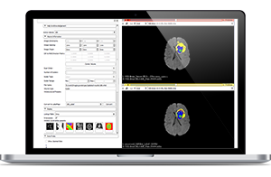
Medical image analysis possibilities for 3D mammography
Tomosynthesis allows for a clearer view of a patient’s breast tissue, yet the recall or misdiagnosis chance still burdens radiologists. Computer-aided diagnosis (CAD) in mammography can lighten this burden by calling for medical imaging software and artificial intelligence to pinpoint even the smallest lesions and attract a specialist’s attention to suspicious regions.
A mammographic CAD system usually consists of several stages, among them:
- Image preprocessing
- Feature extraction and selection
- Segmentation
- Registration
- Classification
The value that computer-aided diagnosis yields is rooted in segmentation, registration and classification. Segmentation stage enables to highlight suspicious areas, permitting more precise lesion location and facilitating lesion margin analysis. Registration stage can serve multiple purposes, such as merging tomosynthetic images with others acquired via ultrasound or MRI to improve diagnostics.
The task of classification is to put out a diagnostic suggestion, such as no signs of tumors, presence of tumors, presence of microcalcifications, etc. Depending on the system’s capabilities, some mammography CADs can distinguish between malignant and benign tumors as well.
Accuracy of mammography CAD systems
As most computer-aided diagnostic solutions are custom and created on demand, the accuracy of each system can vary depending on techniques and approaches used. One of the recently created CAD systems for breast tumor detection was presented in September’s issue of the Applied Soft Computing journal and showed very promising results: 98.69% accuracy, 99.34% sensitivity and 98.26% specificity. This solution employed the Gabor wavelet for feature extraction and Locality Sensitive Discriminant Analysis (LSDA) for data reduction.
In view of such an accuracy rate, the CAD presented is able to ensure diagnostic support without risking to increase the percentage of false positives.
Is mammography CAD evolution worth it?
Answering this question depends on who asks it. Patients are most likely to consider that the alliance of human specialist proficiency and artificial intelligence algorithms will enable early diagnosis without callbacks or additional tests. Insurers may also greet cutting on expenses arising from uncertain diagnosis and related medical errors.
However, the situation gets trickier when it comes to the end users of mammography CAD systems – radiologists. Health specialists are well enough fed up with clogged, rigid and non-functional solutions. They break workflows instead of helping out and take significant time to adopt. Therefore, the rule “out with the old and in with the new” may just not be the case here.
Our suggestion to organizations that plan buying or developing a custom mammography CAD solution is to remember about human-oriented UI, since such complex systems are initially task-focused, leaving convenience aside. However, forgetting about radiologists can turn initial investments in a promising technology into waste only because employees couldn’t figure it out.
If you have anything on your mind regarding tomosynthesis or mammography CAD, we’ll be happy to discuss your point in the comments.
Check opportunities
Medical Image Analysis by ScienceSoft
Become innovative in disease prevention, diagnostics and treatment with efficient medical image analysis.
Check opportunities